During the past 20 years, Dr Eric Daiter has successfully helped thousands of couples that have suffered through the grief and emotional trauma of a pregnancy loss. If you have questions about miscarriage or you just want to find a compassionate infertility specialist to guide you, Dr Eric Daiter would be happy to help (in his Edison, NJ office or on the telephone). It is easy, just call us at 908 226 0250 to set up an appointment (leave a message with your name and number if we are unable to get to the phone and someone will call you back).
Availability
"I always try to be available for my patients since I do understand the pain and frustration associated with fertility problems or endometriosis."
Cost
"I understand that the economy is very tough and insurance companies do not cover a lot of the services that might help you. I always try to minimize your out of pocket cost while encouraging the most successful and effective treatments available."
Need help or have a question?
Uterine Septum 1 View 1
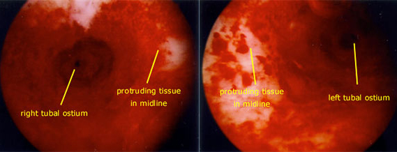
Hysteroscopic views of the uterine cavity (inside lining of the uterus) reveal a wedge shaped defect within this uterine cavity, consisting of tissue that extends from the openings of the right and left fallopian tubes toward the camera (and hysteroscope) that is placed within the uterine cervix (which is the mouth of the uterus that extends into the vaginal vault). Normally, there is no significant amount of uterine tissue between the openings of these fallopian tubes. Laparoscopy provides a panoramic view of the pelvis, including the uterus and the adnexal structures (ovaries and fallopian tubes).
In the upper left hand photo, the opening to the patient's right fallopian tube can be seen as a small dark circle. When viewing this region of the uterine cavity, it appears that you are looking into a cylindrical structure ("like a can"), since there is an excessive amount of tissue to the right of (medial to) the tubal opening.
In the upper right hand photo, the opening of the patient's left fallopian tube can also be seen as a small dark circle. Again, there is a cylindrical appearance to this portion of the uterine cavity, due to an excessive wedge of tissue to the left of (medial to) the tubal opening.