| visit: www.infertilitytutorials.com |
|
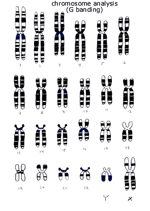
Chromosome Analysis
The DNA of a human cell is packaged into structures known as chromosomes. DNA takes the shape of a double helix and this double helix wraps around small proteins (histones and non histone proteins) within the nucleus of a cell to form chromatin. Chromosomes become visible (due to the condensation of chromatin) using light microscopy (under the microscope) during particular stages (eg, late prophase and metaphase) of cell duplication (mitosis). Human chromosomes are classified by the position of their centromere (primary constriction site) into three types: metacentric (centromere at or near the middle of the chromosome), sub-metacentric (centromere closer to one end), and acrocentric (centromere very close to one end). The blood of healthy individuals does not contain actively dividing cells, so these blood cells are artificially stimulated to divide (undergo mitosis) in order to obtain cells suitable for chromosome analysis. Chromosome preparations from cells that have then been arrested in metaphase are stained and a characteristic banding pattern (of duplicated sister chromosomes) normally appears (black bands represent G bands that are revealed by Giemsa stain, blue bands represent variable regions). Bands on chromosome analysis comprise large segments of DNA and almost always stain multiple different genes. A normal human chromosome complement comprises 23 pairs of chromosomes, a pair of each of 22 autosomes (numbered 1-22) and two sex chromosomes (two X chromosomes for females and one X with one Y chromosome for males). Meiosis is a process that occurs to allow mature eggs or sperm to develop. Meiosis involves a pair of cell divisions during which diploid cells (containing a pair of each of the 23 chromosomes) double their amount of chromosomes and then divide twice so that each resulting cell has only one (rather than 2) of each chromosome. Then, when fertilization occurs the resulting fertilized egg has a total of 23 pairs of chromosomes (one of each of the 23 chromosomes from the egg and the other 23 chromosomes from the sperm). In men, mature sperm are formed in about 74 days. In women, the eggs complete their initial stage (division) of meiosis following the LH surge that triggers ovulation (within about 36 hours). Subsequently, the second stage (division) of meiosis (for an egg) occurs at fertilization (in less than an hour). If the chromosomes do not divide normally during meiosis, nondisjunction (failure of two members of a chromosome pair to separate or disjoin during one of the phases of meiosis) of a specific (pair of) chromosome(s) may result in the mature egg (sperm) having one too many or one too few chromosomes. Chromosome abnormalities occur in about 7% of all conceptions, over 50% of spontaneous abortions (miscarriages), and less than 1% of live-borns. | |||||||||||